Analyze your 3D data with AssayScope


Why choose
AssayScope?
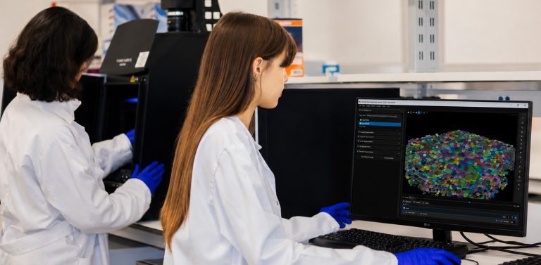
AssayScope, the high-content 3D segmentation and screening software, was designed to simplify the analysis of complex images of 3D cell cultures, organoids, and spheroids. Using cutting-edge artificial intelligence technology, AssayScope allows you to easily extract crucial information and visualize results instantly, without requiring advanced technical expertise. Navigate seamlessly through 3D datasets, segment and analyze cellular structures, and gain meaningful insights in just a few clicks.
Benefit from a cost-effective solution that eliminates the need for expensive equipment or intensive training programs. AssayScope optimizes your workflows and provides powerful analytical tools for 3D cell cultures, saving you time and maximizing your resources.

-
High-precision 3D segmentation
AssayScope offers robust segmentation of nuclei, cells, organoids, and spheroids under real-world conditions, while remaining compatible with a wide range of image qualities, resolutions, and 3D tissue dimensions. This enables users to work with diverse samples. -
3D visualization
With its 3D visualization capabilities, AssayScope provides surface and volumetric renderings of organoids and spheroids, enhancing the visual understanding of these structures. -
Cell and Organoid
Simple and efficient cell and organoid segmentations. Feature extraction (shapes and channels). -
User Interactivity
Link between images and charts for immersive navigation and smooth data exploration. -
Plots and Statistics
Proves all charts and plots adapted to population analysis, z-factor, t-test, pca adapted to segmented 3D imaging.
AssayScope: a powerful and intuitive software designed specifically for biologists and pathologists
AssayScope meets the needs of researchers and biologists working with organoids, spheroids, and 3D tissues by enabling them to:
• Quickly and accurately segment nuclei, cells, and complex 3D structures.
• Analyze large series of images from high-content screening.
• Extract reliable quantitative measurements (size, shape, intensity, distribution, etc.).
• Visualize results in 3D to better understand biological structures.
• Directly link statistical data to source images.
• Identify and isolate cell subpopulations through interactive gating.
• Reduce reliance on lengthy and subjective manual analyses.
• Streamline your image analysis workflow while maintaining the reproducibility of results.
-
Segmentation of 3D nuclei
AssayScope integrates advanced segmentation algorithms based on artificial intelligence, optimized for nuclei, cells, organoids, and spheroids in 3D.
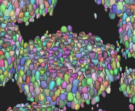
Segmentation of 3D nuclei in organoids
Features
Robust AI algorithms trained on a wide variety of image types and experimental conditions.
Compatibility with different acquisition resolutions and qualities.
Segmentation adapted to simple or complex structures, even with noise or overlaps.
Automated extraction of morphological and intensity measurements.
User experience
Reliable results in just a few clicks, without complex settings.
Immediate visualization of segmented masks in 3D.
Reduction of bias associated with manual segmentations.
Significant time savings in screening and analysis workflows.
3D Nuclei Segmentation Quality
-
Segmentation of 3D cells
3D Cells Segmentation Quality
© Images of organoids courtesy from Furlan and Vincent, CANTHER lab and OrgaRes platform, Univ.Lille, CNRS, Inserm
-
Segmentation of 3D organoids
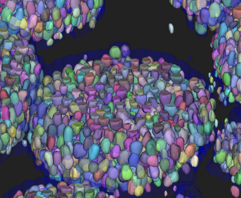
Segmentation of 3D cells and organoids
3D Organoids Segmentation Quality
© Images of organoids courtesy from Furlan and Vincent, CANTHER lab and OrgaRes platform, Univ.Lille, CNRS, Inserm
-
Gating of cell populations with 3D image feedback
AssayScope features an interactive gating tool that allows you to easily select cell subpopulations directly from your analysis plots.
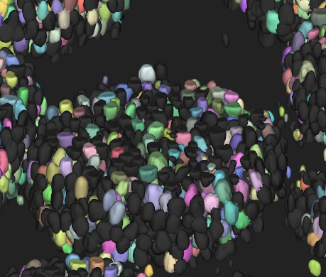
Gating by nucleus size
Features
> Selection on histograms, scatter plots, or population graphs.
> Automatic highlighting of selected cells in the 3D view.
> Dynamic adjustment of thresholds and criteria (size, intensity, shape, etc.).
User experience
> Smooth navigation between statistics and images.
> Immediate understanding of the chosen population’s characteristics.
> Considerable time savings in data exploration and interpretation.
Gating with 3D image feedback
© Images of organoids courtesy from Furlan and Vincent, CANTHER lab and OrgaRes platform, Univ.Lille, CNRS, Inserm
Try AssayScope for free with your own data!
Request a trial versionDiscover our quantitative histological image analysis service
Discover the service