3D Organoid
Image Analysis Services
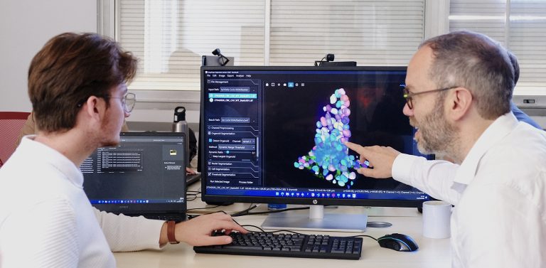
Transform your complex 3D images into clear, actionable data.
See prices


What can we quantify?
QuantaCell extracts robust quantitative data from 3D organoid, spheroid and tissue model images.
- 3D segmentation and reconstruction
Detection of organoids, spheroids, cells, nuclei and relevant 3D structures from image stacks. - Morphological measurements
Quantification of volume, diameter, surface, shape, compactness and structural heterogeneity. - Fluorescence-based quantification
Measurement of marker intensity, expression patterns and signal localization in 3D. - Spatial measurements
Analysis of cell localization, distribution and organization within 3D models. - Condition-based comparisons
Quantitative comparison of image-derived features across treatments, time points or experimental groups.
What can this be used for?
Applications of QuantaCell’s 3D image analysis for organoids, spheroids and 3D tissue models.
- Cellular phenotyping and cell type classification
Characterize cell populations based on morphology, marker expression and spatial localization. - Treatment response assessment
Measure how compounds affect organoid growth, morphology, viability, invasion or degradation. - Detection of biological structures
Identify structures such as rosettes, crypt-like regions, lumens, necrotic cores or other relevant compartments. - Spatial biology in 3D models
Study cell organization, distribution and interactions within organoids, spheroids or patient-derived cultures. - Small-molecule screening
Automate quantitative readouts across multiple compounds, doses or experimental conditions. - Patient-derived organoid analysis
Quantify patient-specific responses in translational research and precision medicine studies.

QuantaCell's commitment
-
Precision
Segmentation validated on diverse samples, even under challenging conditions -
Speed
Fast turnaround times, even for large image volumes -
Flexibility
Custom analyses tailored to your protocols -
Effortless
Ready-to-use results, no training required.
Methodology
1
Choisissez une
de nos option ci-dessous.
Ou envoyez-nous un email à histology@quantacell.com
2
Nous créerons un accès
personnalisé sur notre
serveur web
3
Envoyez-nous une description
des mesures souhaitées et
téléchargez vos images
4
Recevez les résultats et validons-les ensemble !
Pricing
Welcome
0€ /sample
For testing the service!
- Up to 5 samples
- Basic Structures Segmentation (nuclei + cells + organoids)
- Pre-trained AI Models
- Morphology & Signal Intensities Measurements
- Surface & Volume Visualizations
- Data Delivery (.xlsx/.csv + masks TIFF/multipage TIFF)
- 0 Revision
Analyse Standard
From 29,99€ /sample
For complete routine analysis!
- Unlimited. Volume-based pricing*
- Multiple Structures
- Pre-trained AI Models
- Spatial Distribution (& Morphology, Signal Intensities) Measurements
- Surface & Volume Visualizations
- Data Delivery
- 2 Revisions
Analyse avancée
From 59,99€ /sample
For complex / custom research projects!
- Unlimited. Volume-based pricing*
- Multiple Structures
- Custom AI Training & Prediction
- Spatial Distribution (& Morphology, Signal Intensities) Measurements
- Custom Videos (& Surface, Volume) Visualizations
- AI Models & Data Delivery
- Unlimited Revisions
Do you want to analyze your images yourself?
Discover AssayScope software