
Histology & Digital Pathology
Image Analysis Services
QuantaCell featured on Scientist.com

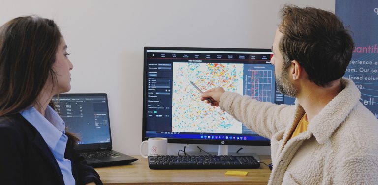



What can we quantify in your histology images?
QuantaCell extracts reliable quantitative data from histology and digital pathology images, from whole-slide tissue structures to single-cell biomarker expression and spatial relationships.
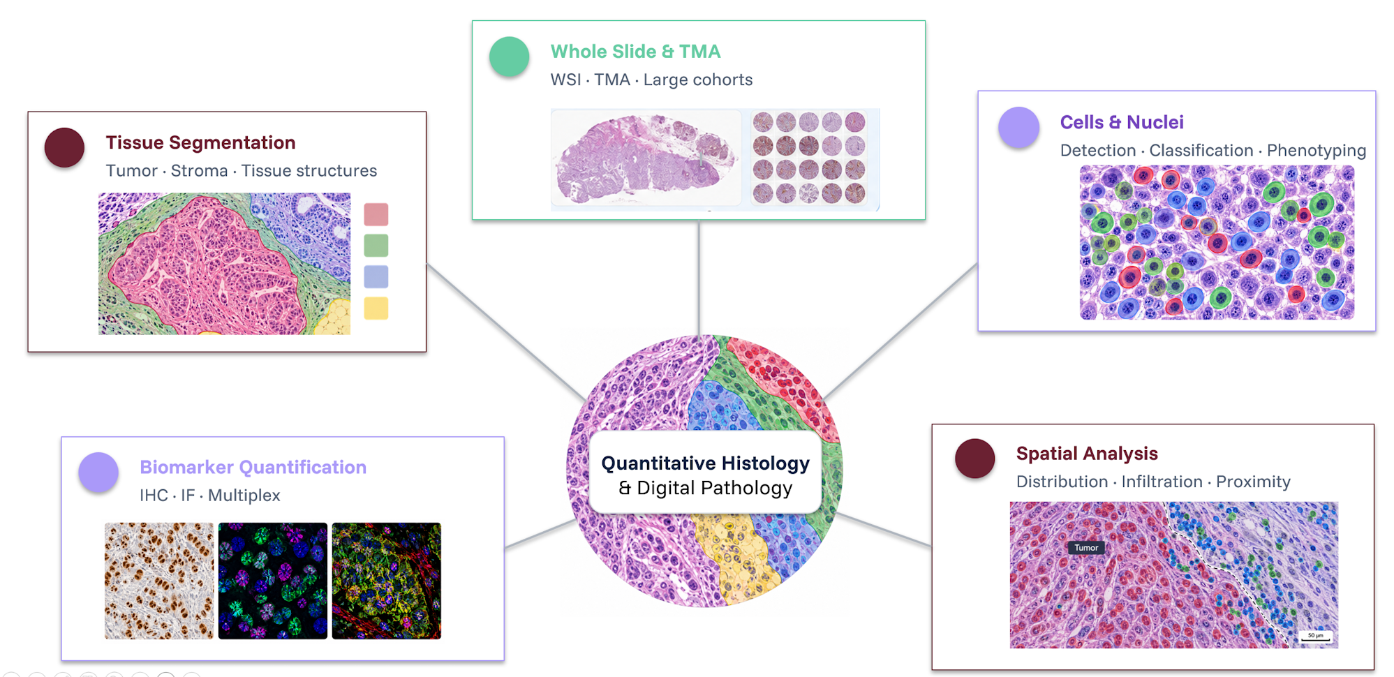
Digital pathology applications
These quantitative readouts support a wide range of digital pathology applications, from biomarker quantification to tissue architecture and spatial analysis.
- Tumor microenvironment analysis
Quantify immune infiltration, tumor–stroma organization and spatial relationships between cell populations and tissue compartments. - Biomarker studies
Measure marker expression, staining intensity, positive cell populations and co-expression across cells and tissue regions. - Tissue morphology & architecture
Analyze tissue compartments, structural organization and morphological changes associated with disease or treatment. - Preclinical & translational research
Compare quantitative histopathology readouts across treatments, experimental models, cohorts or patient samples. - AI-powered digital pathology
Extract reproducible quantitative information from H&E, IHC, immunofluorescence (IF) and multiplex images using advanced image analysis and AI.

QuantaCell's commitment
-
Histology & digital pathology expertise
Our scientists combine expertise in histology, digital pathology, image analysis and AI to develop robust quantitative workflows tailored to your study. -
Fast, reliable quantitative results
Our expertise and established workflows accelerate image analysis while maintaining the flexibility required for complex studies. -
Custom image analysis workflows
We adapt segmentation, classification and quantification methods to your tissue, staining, imaging modality and biological question. -
Scientifically validated results
Intermediate results, segmentation masks and quantitative outputs can be reviewed with your team to ensure biological relevance.
Methodology
1
Share your images.
Send representative images and tell us what you want to quantify.
2
Define the workflow.
We design an analysis workflow tailored to your images and study.
3
Analyze your dataset.
We apply the validated workflow to generate quantitative results.
4
Review the results.
We review the results with you and deliver the quantitative data.
Pricing
Free Feasibility Study
Test up to 5 representative samples to validate feasibility and define the most appropriate analysis workflow.
The feasibility study includes:
- Analysis of up to 5 representative samples
- Feasibility assessment
- Preliminary quantitative results
- Workflow recommendation
Optional service:
- Detailed methodological and analysis report: +€500
Standard Analysis — From €1,500
For studies using established histology and digital pathology analysis workflows.
Typical example: 50 slides or samples
Standard Analysis includes:
- Whole-slide image analysis
- Tissue and/or cell segmentation
- Biomarker quantification
- Quantitative data and visual outputs
- Results review with our team
Optional services:
- Detailed methodological and analysis report: + €500
- Quality tracking & data traceability: + €500
QuantaCell services are also available through
Scientist.com.
Advanced Analysis — From €3,000
For complex studies requiring custom image analysis, advanced quantification or AI model development.
Typical example: 50 slides or samples
Advanced Analysis includes:
- Custom image analysis workflow
- Whole-slide image analysis
- Advanced tissue, cell and spatial analysis
- AI / deep learning model development when required
- Quantitative data and visual outputs
- Results review and workflow optimization
Optional services:
- Detailed methodological and analysis report: + €500
- Quality tracking & data traceability: + €500
QuantaCell services are also available through
Scientist.com.
Do you want to analyze your images yourself?
Discover HistoMetriX software