The AI-Powered organoids, spheroids, and 3D cell cultures quantification software

The most powerful 3D High Content Screening software on the market !

Segment then Analyze !

3D segmentation and high-content screening analysis software to navigate your assay and monitor drug effects.

AssayScope

The software for organoids and 3D cell cultures/tissues to simplify your 3D analyses.

After acquiring your 3D images, you will be able to:

Accurately segment nuclei, cells, organoids, and 3D spheroids,

• Extract key features such as shapes and channels from 3D cell cultures,

• Visualize 3D tissue samples using surface or volumetric rendering,

• Generate and interpret sophisticated graphs and diagrams (population, z-factor, t-test, PCA, …),

• Navigate immersively between 3D images and graphs.

Examples of Applications

• Evaluate the precise impact of drug treatments on cell morphology and viability.

• Understand the formation and behavior of tumor microenvironments by observing and analyzing cell interactions within an organoid or spheroid.

• Develop more targeted and effective treatments by adapting 3D tissue analyses based on the characteristics of a patient’s cells for personalized medicine research.

original 1 XSmallsegm 1 XSmall

Volumetric and surface visualization of a spheroid

“To equip researchers and biologists to tackle the challenges of personalized medicine,

we have developed an AI-enhanced 3D image analysis software.

Why choose AssayScope?

3D SEGMENTATION

HCS ANALYSIS

Robust Nuclei Segmentation

Segment 3D nuclei in real life conditions, compatible with all image qualities, resolutions and dimensions

Visualization

Efficient 3D visualization: surface rendering, volume rendering

Cell and Organoid

Simple and efficient cell and organoid segmentations. Feature extraction (shapes and channels)

User Interactivity

Link between images and charts for immersive navigation and smooth data exploration

Plots and Statistics

Proves all charts and plots adapted to population analysis, z-factor, t-test, pca adapted to segmented 3D imaging

Join the expanding community of researchers

using AssayScope to elevate their 3D analysis

AssayScope meets the needs of researchers and biologists 

working with organoids, spheroids, and 3D tissues by enabling them to:

Quickly and accurately segment nuclei, cells, and complex 3D structures.

• Analyze large series of images from high-content screening.

• Extract reliable quantitative measurements (size, shape, intensity, distribution, etc.).

Visualize results in 3D to better understand biological structures.

• Directly link statistical data to source images.

• Identify and isolate cell subpopulations through interactive gating.

• Reduce reliance on lengthy and subjective manual analyses.

• Streamline your image analysis workflow while maintaining the reproducibility of results.

Segmentation of 3D nuclei

AssayScope integrates advanced segmentation algorithms based on artificial intelligence, optimized for nuclei, cells, organoids, and spheroids in 3D.

Segmentation of 3D nuclei in organoids

Features

  • Robust AI algorithms trained on a wide variety of image types and experimental conditions.


  • Compatibility with different acquisition resolutions and qualities.


  • Segmentation adapted to simple or complex structures, even with noise or overlaps.


  • Automated extraction of morphological and intensity measurements.

User experience

  •  Reliable results in just a few clicks, without complex settings.


  • Immediate visualization of segmented masks in 3D.


  • Reduction of bias associated with manual segmentations.


  • Significant time savings in screening and analysis workflows.

3D Nuclei Segmentation Quality

Segmentation of 3D Cells

3D Cells Segmentation Quality

Segmentation of 3D organoids

Segmentation of 3D cells and organoids

3D Organoids Segmentation Quality

Gating of cell populations with 3D image feedback

AssayScope features an interactive gating tool that allows you to easily select cell subpopulations directly from your analysis plots.

Gating by nucleus size

Features

  • Selection on histograms, scatter plots, or population graphs.


  • Automatic highlighting of selected cells in the 3D view.


  • Dynamic adjustment of thresholds and criteria (size, intensity, shape, etc.).

User experience

  • Smooth navigation between statistics and images.


  • Immediate understanding of the chosen population’s characteristics.


  • Considerable time savings in data exploration and interpretation.

Gating with 3D image feedback

Enhance 3D Analysis with AssayScope

Accelerate your research on organoids, spheroids, and 3D tissues using the power of AI.
Conduct 3D analysis of your results directly! No lengthy or complex training required.

Welcome to AssayScope, the high-content 3D segmentation and screening software designed to simplify the analysis of complex 3D cell culture images, organoids, and spheroids. With the most advanced artificial intelligence technology, AssayScope allows you to easily extract crucial information and visualize results instantly, without requiring extensive technical expertise. Effortlessly navigate through 3D datasets, segment and analyze cellular structures, and gain meaningful insights with just a few clicks.

Enjoy a cost-effective solution without the need for expensive equipment or intensive training programs. AssayScope optimizes your workflows and offers powerful analytical tools for 3D cell cultures, helping you save time and maximize resources.

Let’s explore the key features that make AssayScope the essential solution for your 3D analyses on organoids, spheroids, and 3D tissues. Research use only.


Key Strengths of AssayScope

High-Precision 3D Segmentation

AssayScope offers robust segmentation of nuclei, cells, organoids, and spheroids under real-world conditions, while remaining compatible with a wide range of image qualities, resolutions, and 3D tissue dimensions. This enables users to work with diverse samples.

Visualization

With its 3D visualization capabilities, AssayScope provides surface and volumetric renderings of organoids and spheroids, enhancing the visual understanding of these structures.

Advanced Analysis

Additionally, the graphical and statistical tools provided enable detailed analysis of AI-segmented 3D images, facilitating trend detection and performing specific analyses such as population studies, statistical tests (z-factor, t-test), and principal component analysis (PCA).

Immersive Navigation

One of AssayScope’s strengths is its interactive graphical interface, which creates a direct link between 3D cell culture images and graphs. This feature allows for seamless data exploration, making the user experience not only more enjoyable but also more efficient for analysis and decision-making.

Interested in other software or service?

HistoMetriX

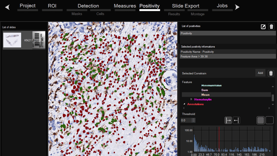
Software for histological samples and TMA

Automatically detects and quantifies your histological slides easily, without requiring bioinformatics expertise. IHC/Fluorescence/Deep learning

QUANTANALYTICS

Services of analysis and characterization

  • Image analysis in patients studies
  • Characterization of drug/compounds effects
  • Automatic detection of rare events

DEEPLEARNING

Services of deep learning generation

  • Creation of precise annotations for training
  • Create powerfull DL models for 2D or 3D
  • Optimize speed and performances

QUANTADEV

Software development for biomedical imaging

  • Customized software solutions with ergonomics UIX
  • AI for automatic analysis
  • Industrialization of analyses in laboratories

Our Last Publications

Our methods and expertise are recognized by the scientific community

through collaborations and peer-reviewed publications

Digitalized organoids

May 2025

With advanced AI and 3D imaging, we reveal organoid structure and dynamics across scales. From nuclei to full organoids, our analysis delivers actionable data for biomedical innovation.

Automated high-speed 3D imaging

July 2022

Our automated platform streamlines 3D organoid culture and imaging, enabling rapid multi-scale analysis. From single cells to entire structures, deep learning quantifies organoid organization with precision.

AI for pulmonary nodule classification

December 2020

By uniting deep learning and medical imaging, we deliver accurate pulmonary nodule detection and classification — achieving 89% sensitivity and 86% specificity to support earlier and more reliable diagnoses.

QuantaCell, Campus Balard, IHU

Contact
+33 (0) 9 83 33 81 90

300 av. du Pr Emile Jeanbrau
34090 Montpellier, France

QuantaCell All rights reserved – 2025