
Projets
et recherche
Nos travaux couvrent l’intelligence artificielle, l’analyse quantitative d’images biomédicales, la pathologie numérique, l’imagerie 3D et la recherche sur les organoïdes.




Nos Collaborations Scientifiques
Chez QuantaCell, nous collaborons dans de nombreux cadres de recherche, notamment des collaborations académiques et hospitalières, des partenariats industriels, des projets ANR (PRC, PRCI et PRCE), des appels à projets régionaux et nationaux, des programmes européens tels que Horizon Europe, EIC et ERC, ainsi que des dispositifs de financement internationaux tels que NIH, NSF et SBIR/STTR.
À l’interface entre sciences du vivant, imagerie, données et logiciel, nous apportons une expertise scientifique, méthodologique et opérationnelle pour transformer des questions biologiques et médicales complexes en workflows d’analyse robustes, exploitables et scientifiquement pertinents.
Vous préparez un projet de recherche ?
Nous serions ravis d’en discuter avec vous dès les premières étapes et d’étudier comment QuantaCell pourrait apporter son expertise à votre projet.
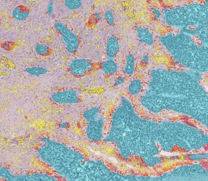
CHU de Montpellier — Anatomopathologie, France
PI : Vanessa Lacheretz Szablewski
Pathologie numérique & histologie quantitative — QuantaCell collabore avec le CHU de Montpellier sur des projets de recherche clinique faisant appel à l’analyse d’images histologiques par IA, à la segmentation tissulaire, à la classification cellulaire et à l’analyse spatiale. Ces collaborations associent expertise en analyse d’images et connaissances anatomopathologiques et cliniques afin d’extraire des informations quantitatives robustes à partir d’images tissulaires complexes.
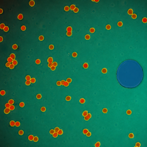
Institut Curie, Paris
PI : Matthieu Piel
Microscopie quantitative & analyse d’images biomédicales — QuantaCell collabore avec l’Institut Curie au développement et à l’application de méthodes avancées d’analyse d’images biomédicales. Notre expertise en segmentation d’images, microscopie quantitative et IA permet de transformer des images de microscopie complexes en données quantitatives reproductibles pour la recherche biologique.
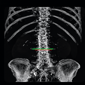
Centre Léon Bérard – Lyon, France
PIs : Pierre-Etienne Heudel, Hugo Crochet
Analyse quantitative d’images en oncologie — QuantaCell apporte son expertise en analyse d’images à des projets de recherche en oncologie portant sur les tissus tumoraux, les biomarqueurs et le microenvironnement tumoral. L’histologie quantitative et l’analyse spatiale permettent de mesurer objectivement l’architecture tissulaire, les phénotypes cellulaires et l’expression des biomarqueurs pour la recherche translationnelle et clinique.
Découvrir le projet sur le site du Centre Léon Bérard →
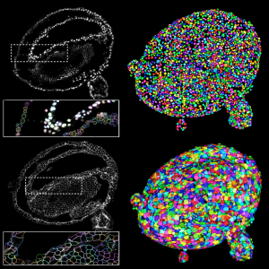
Institute of Mechanobiology, Université de Singapour
PI : Anne Beghin
Analyse d’images 3D d’organoïdes & phénotypage — QuantaCell collabore avec la National University of Singapore sur l’analyse quantitative d’images 3D d’organoïdes. Nos approches combinent segmentation 3D, analyse multi-échelle, phénotypage cellulaire et visualisation afin d’extraire des informations quantitatives à partir de données complexes de microscopie 3D.

IHU Immun4Cure — Montpellier, France
PIs : Jérôme Reboul, Anthony Boureux, Kevin Yauy
Machine learning & analyse de données biomédicales multimodales — QuantaCell apporte son expertise en machine learning et l’analyse de données biomédicales pour intégrer des données biologiques complexes, notamment des données d’imagerie et multi-omiques. Ces approches visent à identifier des signatures quantitatives, des biomarqueurs et des profils biologiques difficiles à mettre en évidence à partir de sources de données analysées séparément.
Découvrir le rapport annuel de l’IHU Immun4Cure →
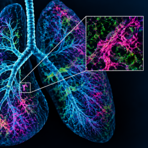
ANR FIRE (Fibrose pulmonaire, imagerie 3D & IA) — Nice France
PI : Chloé Féral (IRCAN)
Fibrose pulmonaire, imagerie 3D & IA — Dans le cadre du projet ANR FIRE, QuantaCell développe des méthodes de deep learning pour l’analyse quantitative 3D d’images de poumon acquises en microscopie light-sheet. Le projet combine segmentation par U-Net 3D, reconstruction des ramifications pulmonaires et détection et quantification de la fibrose, ainsi qu’une application interactive d’annotation 3D pour contribuer à la caractérisation de la fibrose pulmonaire.
Découvrir le projet FIRE sur le site de l’ANR →
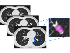
IBM × QuantaCell (IA pour l’imagerie médicale) — Data Challenge JFR 2019
PI : Jean Armand Broyelle
IA pour l’imagerie médicale – QuantaCell a collaboré avec IBM lors du Data Challenge 2019 de la Société Française de Radiologie (JFR), autour du développement de méthodes d’IA pour la détection et la classification de nodules pulmonaires en imagerie scanner 3D. L’équipe IBM–QuantaCell a obtenu le meilleur résultat parmi les dix équipes participantes. Ces travaux ont conduit à la publication scientifique Artificial intelligence solution to classify pulmonary nodules on CT dans Diagnostic and Interventional Imaging (2020).

Nos publications
scientifiques
iScience - Janvier 2026
Human iPSC-derived airway models enable comparative analysis of SARS-CoV-2 infection in healthy and COPD bronchial epithelium
Lisa Morichon, Jitendriya Swain, Nathalie Gros, Amel Nasri, Florent Foisset, Gaetan Galisot, Victor Racine, Said Assou, Arnaud Bourdin, John De Vos, Delphine Muriaux
PMID: 41782833 PMCID: PMC12955582 DOI: 10.1016/j.isci.2026.114741
Keywords: Quantitative image analysis · Airway models · Cell quantification · Fluorescence microscopy · Biomedical image analysis

Nature Methods - Mai 2025
Digitalized organoids: integrated pipeline for high-speed 3D analysis of organoid structures using multilevel segmentation and cellular topology
Nat Methods.2025 May;22, 1343–1354. doi: 10.1038/s41592-025-02685-4. Epub 2025 May 14. Autors : Hui Ting Ong, Esra Karatas, Titouan Poquillon, Gianluca Grenci, Alessandro Furlan, Florian Dilasser, Saburnisha Binte Mohamad Raffi, Damien Blanc, Elise Drimaracci, Dimitri Mikec, Gaetan Galisot, Blake A. Johnson, Albert Zou Liu, Cora Thiel, Oliver Ullrich, OrgaRES Consortium, Victor Racine & Anne Beghin. Keywords : Organoids, Morphology, Biomedical Research

Parasites & Vectors - Mars 2025
Computational histology reveals that concomitant application of insect repellent with sunscreen impairs UV protection in an ex vivo human skin model
Sophie Charrasse, Titouan Poquillon, Charlotte Saint-Omer, Audrey Schunemann, Mylène Weill, Victor Racine, Abdel Aouacheria
PMID: 40038831 PMCID: PMC11881410 DOI: 10.1186/s13071-025-06712-3. Keywords: Computational histology · Histology image analysis · Deep learning · Nuclear morphometry · Skin image analysis · Quantitative pathology
J Clin Invest - Avril 2024
Human NLRC4 expression promotes cancer survival and associates with type I interferon signaling and immune infiltration
23,134(11):e166085. doi: 10.1172/JCI166085. Authors : Charlotte Domblides, Steven Crampton, Hong Liu, Juliet M Bartleson, Annie Nguyen, Claudia Champagne, Emily E Landy, Lindsey Spiker, Christopher Proffitt, Sunil Bhattarai, Anissa P Grawe, Matias Fuentealba Valenzuela, Lydia Lartigue, Isabelle Mahouche, Jeremy Dupaul-Chicoine, Kazuho Nishimura, Félix Lefort, Marie Decraecker, Valérie Velasco, Sonia Netzer, Vincent Pitard, Christian Roy, Isabelle Soubeyran, Victor Racine, Patrick Blanco, Julie Déchanet-Merville, Maya Saleh, Scott W Canna, David Furman, Benjamin Faustin. Keywords: Cancer immunotherapy, Cellular immune response, Immunology, Innate immunity, Oncology.

PLOS ONE, - Mars 2024
Quantitative imaging and semiotic phenotyping of mitochondrial network morphology in live human cells
Sophie Charrasse, Victor Racine, Charlotte Saint-Omer, Titouan Poquillon, Loïc Lionnard, Marine Ledru, Christophe Gonindard, Sandrine Delaunois, Karima Kissa, Richard E Frye, Manuela Pastore, Christelle Reynes, Mathilde Frechet, Hanane Chajra, Abdel Aouacheria
Keywords: Quantitative imaging · Automated image analysis · Live-cell imaging · Morphological profiling · Confocal microscopy · Cellular phenotyping
Front nucl med - Janvier 2024
Automatic deep learning method for third lumbar selection and body composition evaluation on CT scans of cancer patients
10,3:1292676. doi: 10.3389/fnume.2023.1292676. eCollection 2023. Authors : Lidia Delrieu, Damien Blanc, Amine Bouhamama, Fabien Reyal, Frank Pilleul, Victor Racine, Anne Sophie Hamy, Hugo Crochet, Timothée Marchal, Pierre Etienne Heudel. Keywords: body composition, cancer; computed tomography, deep learning, sarcopenia.
Comput Struct Biotechnol J - Novembre 2023
Quantitative assessment of mitochondrial morphology relevant for studies on cellular health and environmental toxicity
Sophie Charrasse, Titouan Poquillon, Charlotte Saint-Omer, Manuela Pastore, Benoit Bordignon, Richard E Frye, Christelle Reynes, Victor Racine, Abdel Aouacheria
Keywords: High-content screening · Quantitative imaging · Automated image analysis · Morphological profiling · Live-cell imaging · Toxicology
Nature Methods - Juillet 2022
Automated high-speed 3D imaging of organoid cultures with multi-scale phenotypic quantification
19(7):881-892. doi: 10.1038/s41592-022-01508-0. Epub 2022 Jun 13.Autors : Anne Beghin, Gianluca Grenci, Geetika Sahni, Su Guo, Harini Rajendiran, Tom Delaire, Saburnisha Binte Mohamad Raffi, Damien Blanc, Richard de Mets, Hui Ting Ong, Xareni Galindo, Anais Monet, Vidhyalakshmi Acharya, Victor Racine, Florian Levet, Remi Galland, Jean-Baptiste Sibarita, Virgile Viasnoff. Keywords : 3D imaging, Deep Learning, Quantification.

Diagn Interv Imaging - Décembre 2020
Artificial intelligence solution to classify pulmonary nodules on CT
101(12):803-810. Autors : D Blanc, V Racine, A Khalil, M Deloche, J-A Broyelle, I Hammouamri, E Sinitambirivoutin, M Fiammante, E Verdier, T Besson, A Sadate, M Lederlin, F Laurent, G Chassagnon, G Ferretti, Y Diascorn, P-Y Brillet, Lucie Cassagnes, C Caramella, A Loubet, N Abassebay, P Cuingnet, M Ohana, J Behr, A Ginzac, H Veyssiere, X Durando, I Bousaïd, N Lassau, J Brehant. Keywords : Deep learning, Retina-UNET, 3D U-Net, PyTorch

Transl Psychiatry - Octobre 2020
Mitochondrial morphology is associated with respiratory chain uncoupling in autism spectrum disorder
11(1):527. Autors : RE Frye, L Lionnard, I Singh, MA Karim, H Chajra, M Frechet, K Kissa, V Racine, A Ammanamanchi, PJ McCarty, L Delhey, M Tippett, S Rose, A Aouacheria. Keywords : Cell segmentation, nuclei segmentation, statistics, AssayScope
Methods Cell Biol - Janvier 2017
Fluorescence eXclusion Measurement of volume in live cell
Auteurs : Cadart C, Zlotek-Zlotkiewicz E, Venkova L, Thouvenin O, Racine V, Le Berre M, Monnier S, Piel M. Keywords : Mass analysis. Biophysics, Microfluidcs
Nature Commun - Juin 2014
Synaptic recruitment of gephyrin regulates surface GABAA receptor dynamics for the expression of inhibitory LTP
Auteurs :Petrini EM, Ravasenga T, Hausrat TJ, Iurilli G, Olcese U, Racine V, Sibarita JB, Jacob TC, Moss SJ, Benfenati F, Medini P, Kneussel M, Barberis A. Keywords : Neuroscience, dynamique des récepteurs, suivi dans le temps, segmentation
Optics Express - Janvier 2012
Wavelet analysis for single molecule localization microscopy
20(3):2081-95. PDF Auteurs :Izeddin I, Boulanger J, Racine V, Specht CG, Kechkar A, Nair D, Triller A, Choquet D, Dahan M, Sibarita JB. Keywords : TIRF/PALM, SPT, détection moléculaire, imagerie avancée, ondelettes, fit gaussien, traitement des images
Molecular Systems Biology - Janvier 2012
RNAi screening reveals a large signaling network controlling the Golgi apparatus in human cells
Auteurs :Chia J, Goh G, Racine V, Ng S, Kumar P, Bard F. Keywords : Criblage phénotypique, structure cellulaire, siRNA, machine learning, statistiques
Nature - Mai 2007
Experimental and theoretical study of mitotic spindle orientation
447(7143):493-6. Auteurs :Théry M, Jiménez-Dalmaroni A, Racine V, Bornens M, Jülicher F. Keywords : Cellule unique, détection et segmentation de compartiments, adhésion, microscopie automatisée, visualisation des données, modèle théorique, cytosquelette
De la recherche à l’analyse d’images appliquée
Nos travaux scientifiques contribuent directement au développement des méthodes et technologies d’analyse d’images de QuantaCell. De l’histologie et la pathologie numérique aux organoïdes 3D, à la microscopie quantitative et à l’IA, cette expertise issue de la recherche nous permet de développer des workflows d’analyse robustes pour répondre à des problématiques biologiques complexes.
Vous souhaitez plus d’informations ?
Contactez-nous